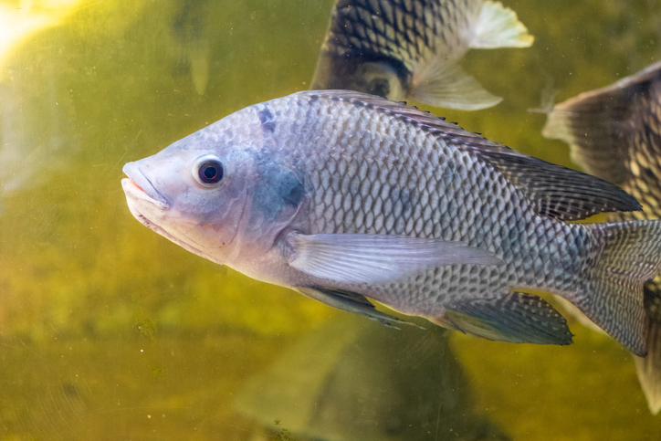
Tilapia skin to reconstruct child’s fingers
Cleaning, freeze-drying and sterilization by irradiation is a combination of processes that have now been used for a few decades to prepare human replacement tissues in surgical procedures. One example is bone grafts taken from corpses used to fill holes caused by trauma, cancer, or other diseases in bones. Over the years the sources and types of biomaterials and the purposes have diversified. A recent example comes from Brazil where tilapa fish skin is used.
The Apert syndrome is a rare genetic defect that affects one in every 70,000 born. One of the symptoms is syndactyly, the webbing of fingers and toes. in 2018 a team in Brazil developed a protocol for the separation of fingers from Apert carriers to maximize the movement and function of the hands. Surgical separation of the fingers leaves a region skinless and requires skin from the child’s own abdomen to be grafted there to reconstitute the tissue . Then the Federal University of Ceará (UFC) and The Sobrapar Hospital in Campinas (São Paulo region) developed an alternative where pieces of tilapia skin are used to improve the process of reconstitution of the fingers and to optimize recovery. The skin of the fish is formed in a fabric, frreeze-dried, vacuum packed and gamma sterilized at the facility of our member IPEN in São Paulo.
The first surgeries occurred in September 2021. The technique has a number of advantages: reduction in the time of surgery, lower morbidity of grafted tissue and better grip (or adherence) of the human skin graft in the region where the fingers are separated. The use of biomaterial also decreased the number of dressings by 50%, caused relief in postoperative pain and resulted in lower treatment costs.
The tilapia skin thus evolved from a dressing to treat burns and injuries to a biomaterial that prepares the skin to receive grafts, either in gynecology – in surgeries for vaginal reconstruction – or in the treatment of Apert. According to Dr Edmar Maciel, surgeon at the Burns Support Institute (IAQ) in Fortaleza: “In deep wounds, the collagen of the tilapia skin is absorbed and integrated into the wound bed. This collagen creates a dermal matrix to receive the graft with the patient’s own skin, something that happens 10 days after the application of fish tissue“. When the tilapia skin is removed, the wound is prepared to receive pieces of skin thinner than abdominal tissue, usually used in conventional procedures. The surgeon adds “The new technique facilitates the integration of structurally simpler tissues – such as skin removed from the patient’s scalp or forearm – with the wound bed. This increases the success of the skin handle and prevents a large scar on the belly“.
